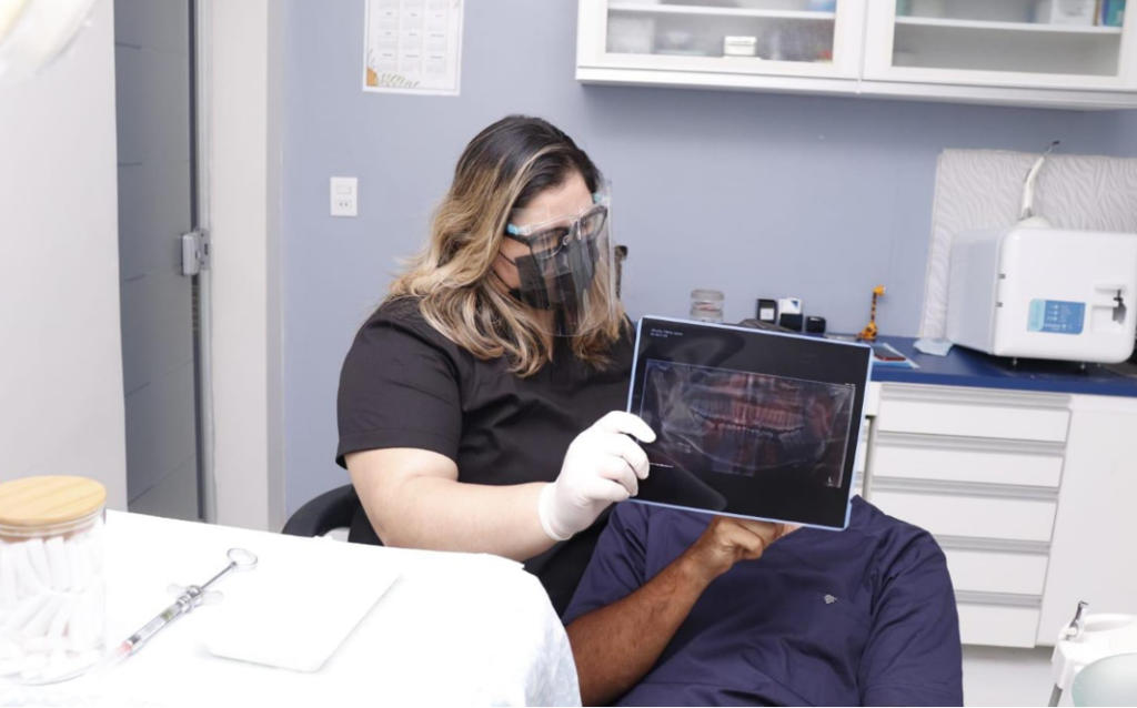
X-rays are a fundamental tool in dentistry, as they allow the dentist to obtain a clear and detailed view of the internal structures of the teeth and jawbones. There are several types of intra- and extra-oral X-rays, including:
Periapical X-rays: These images focus on one or two teeth and show the crown, root and surrounding bone. They are useful for detecting cavities, infections and root problems.
Panoramic X-rays: These provide a complete view of the mouth, including all the teeth, jawbones and surrounding structures. They are especially useful for planning orthodontic treatments and for assessing the position of impacted or impacted teeth.
IMPORTANCE OF X-RAYS
X-rays are essential for an accurate diagnosis. They allow dentists to identify problems that are not visible in a clinical evaluation, such as hidden cavities, abscesses, cysts and periodontal disease. Furthermore, they are essential for monitoring treatments, such as evaluating the effectiveness of a root canal treatment, observing tooth eruption in children, correctly planning orthodontic treatment, planning tooth extractions, etc. Failure to use imaging studies can lead to misdiagnosis and treatment failures.
